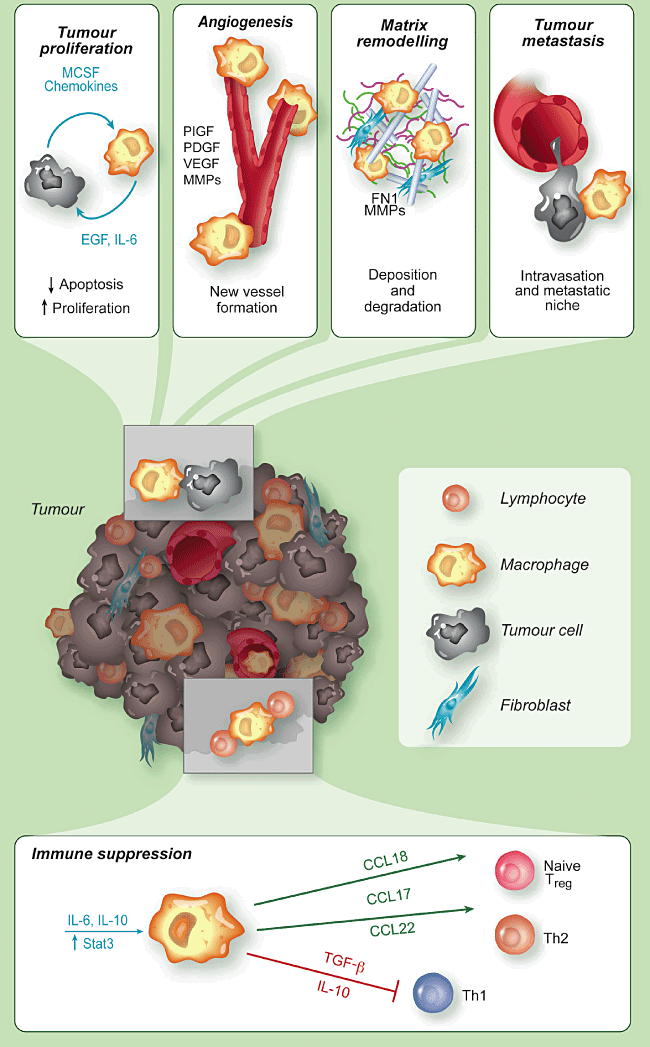
RECOMMENDED CANCER, STARVING DIET, MACROPHAGES


Home
3-bromopyruvate, starving cancer
90% CHEMO SUCKS--BMJ REVIEW, 2016
pharma and chemo--sucks
Cancer basics and starving cancer--jk
Starving cancer by fasting and ketogenic diet, a review
Cancer as a metabolic disease, starving cancer--Seyfried, 2014
Highlites of Seyfried 2014 Plus 2 more articles
Warburg effect--Dr. Fung Blog-2-18
Vitamin C prolongs life metastatic patients
Ketogenic diet starves cancer, Seyfried Journal 2007
Role of Macrophages in metastatic cancer
Metabolic pathways and cancer growth--2008 review
Glutimate cancer treatments
Glutamine and cancer-2001 review
Blocking Glutimate metabolism by cancer
Ketogenic diet starves cancer, known as Warburg effect, 1924
Otto Warburg's article plus study of Warburg effect
Mega Vitamin C slows cancer invasion, Pauling trial

Role of Macrophages in metastatic cancer


Like so much of funding driven science, look for a drug to treat rather than prevent or starve cancer, thus using drugs to target macrophages to improve results of chemotherapy.  As stated in my article on cancer, this approach at best extends life a few month and doesn’t produce a cure—with the exception of about 4 cancer where chemo can cure or cause long-term remissions.  The problem with a drug that targeting TAM, is that it will target macrophages, it will target all M2 macrophages and very possible other immune cells with consequences that likely will limit the therapy to short term with and produce significant side effects. 

^^^^^^^


Tumour-associated macrophages: undisputed stars of the inflammatory tumour microenvironment


·         P. Allavena,  A. Mantovani


http://onlinelibrary.wiley.com/doi/10.1111/j.1365-2249.2011.04515.x/full   Clinical and experimental Immunology Volume 167, Issue 2, February 2012 ,  Pages 195–205


Summary:


Mononuclear phagocytes are cells of the innate immunity that defend the host against harmful pathogens and heal tissues after injury. Contrary to expectations, in malignancies, tumour-associated macrophages (TAM) promote disease progression by supporting cancer cell survival, proliferation and invasion. TAM and related myeloid cells [Tie2+ monocytes and myeloid-derived suppressor cells (MDSC)] also promote tumour angiogenesis and suppress adaptive immune responses. These divergent biological activities are mediated by macrophages/myeloid cells with distinct functional polarization, which are ultimately dictated by microenvironmental cues. Clinical and experimental evidence has shown that cancer tissues with high infiltration of TAM are associated with poor patient prognosis and resistance to therapies. Targeting of macrophages in tumours is considered a promising therapeutic strategy: depletion of TAM or their ‘re-education’ as anti-tumour effectors is under clinical investigation and will hopefully contribute to the success of conventional anti-cancer treatments.   [myelocyte is a young cell of the granulocytic series, occurring normally in bone marrow (can be found in circulating blood when caused by certain diseases) wiki.]


Introduction:


The complex relationship between tumours and the immune system has long been studied. Cancer cells express tumour-associated antigens able to trigger the host immune response [1–3]. Indeed, in recent decades there has been growing evidence that cells of the adaptive immunity are present at tumour sites and are usually associated with more favourable prognosis. The best evidence is in human colorectal cancer, where CD3+CD8RO+ lymphocytes are clearly associated with longer disease-free and -specific survival [4,5]. Also in other human tumours, such as ovary, melanoma and breast, the density of T cells is usually correlated with a more favourable outcome [6–9]. In marked contrast, in the majority of cancers, cells of the innate immunity, especially macrophages, most frequently favour tumour progression.  Tumour-associated macrophages (TAM) are recruited early at tumour sites where they most frequently display pro-tumour functions, such as activation of the neo-angiogenic switch, the secretion of soluble factors that support tumour cell resistance to apoptotic stimuli and stimulate the proliferation and invasion of malignant cells. TAM have been also associated with the suppression of adaptive immunity [10–15].


Thus, in a simplified scheme, adaptive immunity is usually protective and limits tumour progression, while innate immunity favours disease development. However, biological mechanisms are more complex: recent evidence has been provided that components of adaptive immunity [e.g. interleukin (IL)-4-producing CD4 T cells and antibody-producing B cells] may activate innate immune cells in a pro-tumour manner [14,16]. Therefore, the dynamic interplay between innate and adaptive immunity is of paramount importance in the outcome of tumour progression or regression. A further level of complexity is dictated by the multi-faceted functional phenotypes of myeloid cells, especially macrophages, which allow them to have either anti-tumour or pro-tumour activities.


In this review we will summarize the current view on the roles of TAM in tumours and the available strategies to target or exploit them for anti-cancer therapies.


Macrophage heterogeneity


Macrophages are versatile cells that are capable of displaying different functional activities, some of which are antagonistic: they can be immune-stimulatory or immune suppressive, and either promote or restrain inflammation [13,17–22]. This functional plasticity is regulated by local cues to which the macrophages respond. For instance, during bacterial infections macrophages first orchestrate the acute inflammatory response to eliminate the invading pathogens; at later times they transform into scavengers of tissue debris; further on they trigger the proliferative phase of healing by releasing a variety of growth factors and cytokines which recruit and activate fibroblasts and new vessel[20,23].


Macrophage heterogeneity has been simplified in the macrophage polarization concept where the two extreme phenotypes, the M1 and M2 macrophages, have distinct features [17,24–28], as depicted in Fig. 1. M1 or classically activated macrophages are stimulated by bacterial products and T helper type 1 (Th1) cytokines [e.g. interferon (IFN)-γ]; they are potent effectors that produce inflammatory and immune-stimulating cytokines to elicit the adaptive immune response, secrete reactive oxygen species (ROS) and nitrogen intermediates and may have cytotoxic activity to transformed cells. M2 or alternatively activated macrophages differentiate in microenvironments rich in Th2 cytokines (e.g. IL-4, IL-13); they have high scavenging activity, produce several growth factors that activate the process of tissue repair and suppress adaptive immune responses [15,29,30].


monocyte-macrophage-cancer-promoters.gif


Figure 1.


Macrophage polarization is modulated by microenvironmental signals. T helper type 1 (Th1) cytokines [e.g. interferon (IFN-γ)] and Toll-like receptor (TLR) ligands [e.g. lipopolysaccharide (LPS)] promote M1 macrophages which elicit Th1 immune responses and fight intracellular pathogens. Th1 cells produce IFN-γ, which further sustains M1 polarization. Th2 cytokines [e.g. interleukin (IL)-4, IL-13], IL-10 and glucocorticoids promote M2 macrophages which block Th1 immune responses and promote wound healing, scavenging of damaged tissues and angiogenesis. Th2 cells produce IL-4, which further sustains M2 polarization.


Macrophages infiltrate neoplastic lesions from the early stages of tumorigenesis and usually precede other leucocytes (e.g. lymphocytes) having potential anti-tumour function [31]. Tumour and stromal cells secrete a number of diverse chemo-attractants that recruit blood circulating monocytes. For instance, the chemokine CCL2 was discovered as a tumour-derived factor inducing chemotaxis in monocytes [32,33]. Once in tumours, monocytes differentiate to macrophages primarily because of the presence of macrophage colony-stimulating factor (M-CSF), produced by tumour cells. M-CSF production in human tumours correlates with poor prognosis in ovarian, breast and endometrial cancer [34,35]. Conditioned by the tumour milieu, monocytes differentiate as tumour-educated macrophages and acquire properties of immune-suppressive and pro-tumoural effectors.


In established tumours, TAM resemble M2-like macrophages [36–40]. While M2-related activities are of extreme importance during wound healing to return to the homeostatic state, in the context of a growing tumour they may favour disease progression [11,12,36,39,41–43].


In molecular profiling studies, murine [family of rodents including mice and rats} TAM display hallmarks of M2 macrophages: arginase-I, YM1, FIZZ1, MGL2, vascular endothelial growth factor (VEGF), osteopontin and matrix metalloproteinases (MMPs), as well as an immunosuppressive phenotype: high IL-10, transforming growth factor (TGF-β) and low IL-12, reactive nitrogen intermediates (RNI) and major histocompatibility complex (MHC) II, which correlate functionally to reduced cytotoxicity and antigen-presenting capacity [44–46].


Similar findings were found in human TAM from ovarian cancer patients [47]. We compared the expression of up-regulated genes in human TAM with the profiling of in-vitro-polarized M1 and M2 macrophages. Some up-regulated genes (e.g. osteopontin, fibronectin, scavenger and mannose receptors) were up-regulated similarly in TAM and in M2 macrophages. Indeed, using principal component analysis, the global profiling of TAM fell much closer to that of M2-polarized macrophages [40].  In fact, TAM heterogeneity is starting to emerge, probably depending on the tumour type and micro-environmental cues [39,48]. Notably, murine TAM from fibrosarcoma also showed the expression of typical M1 factors such as IFN-inducible chemokines (CCL5, CXCL9, CXCL10, CXCL16) [44,49].


In fact, TAM heterogeneity is starting to emerge, probably depending on the tumour type and micro-environmental cues [39,48]. Notably, murine TAM from fibrosarcoma also showed the expression of typical M1 factors such as IFN-inducible chemokines (CCL5, CXCL9, CXCL10, CXCL16) [44,49].


TAM as promoters of cancer-related inflammation


Conditions of persistent inflammation in tissues predispose to carcinogenesis and, in established malignancies, accelerate tumour development [42,50–54]. Cancer-related inflammation is now recognized as a hallmark of cancer [55,56]. Macrophages are key initiators of the subtle chronic inflammation present in the tumour microenvironment, as they are major producers of inflammatory mediators. Several experimental studies in inflammation-induced murine tumour models have demonstrated the requirement of nuclear factor (NF)-κB activation in TAM for tumour promotion [46,54,57–60].  Within the tumour context the transcription factor NF-κB can be triggered in macrophages by factors released by necrotic tissues [e.g. high-mobility group protein 1 (HMGB1) degraded matrix proteins] and by inflammatory cytokines [e.g. tumour necrosis factor (TNF)] produced by neoplastic cells [61]. Activated TAM, in turn, produce cytokines (IL-6, TNF) and chemokines, which perpetuate and amplify the inflammatory cascade [42].


            The primary inflammatory cytokine TNF, produced by immune cells but also by malignant and stromal cells, is an activating mediator and at low concentrations sustains the growth of tumour cells and blood vessels. TNF is also associated with increased release of chemokines (CCL2, CXCL8, CXCL12) and activation of matrix degrading enzymes.  [61].


IL-6 is a key growth-regulating and anti-apoptotic cytokine, having tumour-inducing activities on both malignant and stromal cells. In recent years, the involvement of IL-6 in cancer has been closely investigated, highlighting NF-kB as a link. In mouse models of colitis, IL-6 is produced mainly by macrophages in response to intestinal injury and in an NF-kB-dependent manner. Inhibition of NF-kB in these cells resulted in reduced tumour growth, which was attributed to decreased IL-6 production by TAM [57,62,63]. In murine models of hepatocellular carcinoma, NF-kB inhibition in liver macrophages (Küpffer cells) resulted in marked delay of tumour onset [64,65].


Further, in tumour areas of low oxygen tension, where TAM usually accumulate, hypoxia induces a hypoxia-induced factor (HIF)-1alpha-mediated transcriptional programme, which includes also the activation of NF-kB in macrophages [66,67]. Collectively, these studies clearly emphasize the essential requirement of NF-kB activation in TAM for maintaining the inflammatory circuit(s) that promote tumour growth.


[Wikipedia : “NF-κB (nuclear factor kappa-light-chain-enhancer of activated B cells) is a protein complex that controls transcription of DNA, cytokine production and cell survival. NF-κB is found in almost all animal cell types and is involved in cellular responses to stimuli such as stress, cytokinesfree radicalsheavy metalsultraviolet irradiation, oxidized LDL, and bacterial or viral antigens.[1][2][3][4][5]  NF-κB plays a key role in regulating the immune response to infection (κ light chains are critical components of immunoglobulins). Incorrect regulation of NF-κB has been linked to cancer, inflammatory and autoimmune diseases, septic shock, viral infection, and improper immune development. NF-κB has also been implicated in processes of synaptic plasticity and memory.[6][7][8][9][10][11]  All proteins of the NF-κB family share a Rel homology domain in their N-terminus. here are five proteins in the mammalian NF-κB family:[17]  This allows NF-κB to be a first responder to harmful cellular stimuli. Known inducers of NF-κB activity are highly variable and include reactive oxygen species (ROS), tumor necrosis factor alpha (TNFα), interleukin 1-beta (IL-1β), bacterial lipopolysaccharides (LPS), isoproterenol, cocaine, and ionizing radiation.[21]  In unstimulated cells, the NF-κB dimers are sequestered in the cytoplasm by a family of inhibitors, called IκBs (Inhibitor of κB), which are proteins that contain multiple copies of a sequence called ankyrin repeats. NF-κB is widely used by eukaryotic cells as a regulator of genes that control cell proliferation and cell survival. As such, many different types of human tumors have misregulated NF-κB: that is, NF-κB is constitutively active. Active NF-κB turns on the expression of genes that keep the cell proliferating and protect the cell from conditions that would otherwise cause it to die via apoptosis.  Data have also shown that NF-κB activity enhances tumor cell sensitivity to apoptosis and senescence. herefore, NF-κB promotes Fas-mediated apoptosis in cancer cells, and thus inhibition of NF-κB may suppress Fas-mediated apoptosis to impair host immune cell-mediated tumor suppression.” https://en.wikipedia.org/wiki/NF-%CE%BAB]


IL-6 activates the signal transducer and activator of transcription 3 (STAT3) pathway, another crucial mediator of the cancer-related inflammation. In tumour cells STAT3 induces the expression of genes important for cell cycle progression [such as cyclin D and proliferating cell nuclear antigen (PCNA)] and suppression of apoptosis (Bcl-XL, Bcl-2 and Mcl-1) [53,68,69]. In a mouse genetic model of pancreatic cancer, STAT3 activation was contributed mainly by macrophage-released IL-6; ablation of IL-6 production or STAT3 activation resulted in decreased carcinogenesis and inflammatory cell infiltration [70,71].


Chemokines and their receptors are key players in cancer-related inflammation, and TAM are a rich source of different inflammatory chemokines. The CXC chemokines bearing the ELR motif (e.g. CXCL1, CXCL2 and the most popular CXCL8) have potent angiogenic function and activate the neo-angiogenic switch [72,73]. CCL2 is highly produced by TAM and amplifies the recruitment of myeloid cells within tumours. Further, it may play an important role in the regulation of angiogenesis [72,74].


During the last decade there has been recognition that degraded/proteolytic fragments of extracellular matrix (ECM) molecules, or their aberrant expression, can sustain the activation of inflammatory cells and contribute to fuel inflammation at tumour sites. A cryptic peptide of laminin-10, a prominent component of basement membranes, is chemotactic for neutrophils and macrophages and induces the up-regulation of TNF, chemokines and MMP-9 [75]; versican activates Toll-like receptor (TLR)-2 and TLR-6 on TAM and stimulates the expression of inflammatory genes [76], while hyaluronan fragments trigger through TLR-4, TLR-2 and the CD44 receptor [77].


Pro-tumour functions of TAM


TAM influence fundamental aspects of tumour biology, as shown in Fig. 2. Among the well-documented pro-tumour functions of TAM is the production of trophic and activating factors for tumour and stromal cells [e.g. endothelial growth factor (EGF), fibroblast growth factor (FGF), VEGF, platelet-derived growth factor (PDGF), TGF-β]. These growth factors directly promote the proliferation of tumour cells and increase resistance to apoptotic stimuli [42,78–80]. TAM are also a major source of proteolytic enzymes that degrade the ECM, thus favouring the release of matrix-bound growth factors [42,81]. As mentioned above, IL-6 released by TAM plays a key role in sustaining the survival and proliferation of malignant cells in tumours of epithelial and haematopoietic origin [63,70,71,82,83].


Illustration here


pro-tumor-function-macrophage.gif


Figure 2.


Pro-tumour functions of tumour-associated macrophages (TAM). TAM promote the survival of neoplastic cells from apoptotic stimuli and their proliferation, by producing several growth factors and cytokines [e.g. epithelial growth factor (EGF), interleukin (IL)-6], and the tumour angiogenesis, via vascular endothelial growth factor (VEGF), matrix metalloproteinases (MMPs) and other angiogenic factors. TAM have an intense proteolityic activity and degrade the extracellular matrix, but also produce matrix proteins, such as fibronectin (FN1). They favour tumour cell intravasation and dissemination to distant sites. TAM have immune suppressive functions by producing IL-10 and transforming growth factor (TGF-β) which suppress T helper type 1 (Th1) lymphocytes, and by secreting chemokines (e.g. CCL17, CCL18, CCL22) which recruit lymphoid cells devoid of cytotoxic activity (Th2, naive lymphocytes) or having suppressive functions [regulatory T cells (Treg)].


TAM are key effectors of the ‘angiogenic switch’ where the balance between pro- and anti-angiogenic factors, commonly present in tissues, tilts towards a pro-angiogenic outcome [84–87]. In hypoxic conditions the transcription factor HIF-1alpha induces in TAM the production of VEGF and the angiogenic chemokine CXCL8 [88].


In addition, a unique subset of monocytes has been identified recently in tumours by the expression of the angiopoietin (Ang) receptor Tie-2, named Tie-2 expressing monocytes (TEMs) [89] Thus, TAM and related myeloid cells directly produce angiogenic factors and are also a major source of proteolytic enzymes which mobilize VEGF from extracellular matrix stores, indirectly sustaining tumour angiogenesis [90].
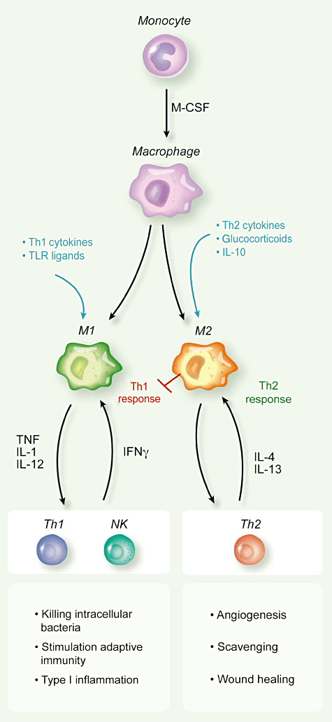

It is emerging that the expression pattern of genes that regulate iron homeostasis is distinct in polarized macrophages. For instance, M1 macrophages showed ferroportin repression and H ferritin induction, thus favouring iron sequestration, whereas M2 macrophages had an inverse expression profile (ferroportin up-regulation and down-regulation of H ferritin and haem oxygenase) and enhanced iron release [91]. Therefore, M1 cells mediate iron retention to control pathogen expansion during the acute phase of inflammation, while M2 cells donate iron that is important to tissue repair in the resolution phase. Interestingly, haem oxygenase 1 is highly expressed in TAM; it is tempting to speculate that the increased iron availability in the tumour microenvironment might represent a previously unknown mechanism that underlies the tumour-promoting activity of TAM [92].


TAM are probably the most active contributors to the incessant matrix remodelling present within tumours, as they produce several MMPs and other proteolytic enzymes [93]. Tumour cells exploit the ECM degradation mediated by TAM to invade locally, penetrate into vessels and disseminate to give distant metastasis [94]. TAM aiding cancer cell invasion have been visualized directly in experimental tumours in vivo by multi-photon microscopy: by using fluorescently labelled cells, Wyckoff and colleagues showed that tumour cell intravasation occurs next to perivascular macrophages in mammary tumours [94,95]. Further, it has been shown recently that cathepsin protease activity, by IL-4-stimulated TAM, promotes tumour invasion [96]. IL-4 is produced by tumour-infiltrating CD4 T cells and there is mounting evidence of its relevance in the polarization of macrophages with pro-tumour functions [14,16]. The chemokine CCL18 produced by TAM has been shown recently to play a critical role in promoting breast cancer invasiveness by activating tumour cell adherence to ECM [97].


We found recently that human TAM and in-vitro tumour-conditioned macrophages express high levels of the migration stimulation factor (MSF) [40], a truncated isoform of fibronectin [98]. Macrophage-secreted MSF displays potent chemotactic activity to tumour cells in vitro [40], confirming that the pro-invasive phenotype of cancer cells is modulated by macrophage products released in the tumour microenvironment.


Further support to the concept of a reciprocal interaction between tumour cells and TAM was provided by a recent paper where SNAIL-expressing keratinocytes became locally invasive after macrophage recruitment elicited by M-CSF [99].


Tumour macrophages have the ability to suppress the adaptive immune response, thus contributing directly to the phenomenon of immune evasion of cancer [1]. TAM are poor antigen-presenting cells, have defective IL-12 secretion [100], produce IL-10 and TGF-β and inhibit T cell proliferation [27,36,101]. At least some of these immune-suppressive activities of TAM are mediated by over-activation of the transcription factor STAT3. In immune cells STAT3 enables suppression of tumour immunity by opposing STAT1-regulated Th1 anti-tumour immune responses and promoting the differentiation of immature myeloid cells with suppressive activity [102].


Myeloid-derived suppressor cells (MDSC), identified in tissues and lymphoid organs of tumour-bearing hosts, contribute to tumour-induced immune suppression [101,103–105]. These cells share properties and gene expression profiles with M2-polarized TAM, yet also display distinct features [106]. MDSC use two enzymes involved in the arginine metabolism to control T cell response: inducible nitric oxide synthase (NOS2) and arginase (Arg1), which deplete the milieu of arginine, causing peroxinitrite generation and T cell apoptosis [107].


The immune-suppressive activity of TAM is also exerted indirectly by their release of chemokines (e.g. CCL17 and CCL22) that preferentially attract Th1, Th2 lymphocytes and regulatory T cells (Treg), devoid of cytotoxic functions [72]. The chemokine CCL18, produced abundantly by TAM from human ovarian carcinoma [108], recruits naive T cells, which eventually turn into anergic cells within a microenvironment dominated by M2 macrophages and immature DC [109,110].


In line with the above experimental evidence, in the majority of human tumours high numbers of infiltrating TAM have been associated significantly with advanced tumours and poor patient prognosis [11,15,42,111]. There are, however, notable exceptions to this pro-tumour phenotype, probably dictated by TAM functional polarization. One such exception is human colorectal cancer, where some studies have reported that TAM density is associated with better prognosis [112–114]. The localization of TAM within colorectal cancers appears to be of primary importance: the number of peritumoural macrophages with high expression of co-stimulatory molecules (CD80 and CD86), but not of those within the cancer stroma, was associated with improved disease-free survival [115,116].


Specific TAM subsets identified by surface markers may have predictive values: in lung adenocarcinoma, the number of TAM CD204+ (scavenger receptor) showed a strong association with poor outcome, while the total CD68+ population did not [117].


Macrophage-related gene signatures have been identified in human tumours such as ovarian and breast cancer, soft tissue sarcoma and follicular B lymphoma [118–121]; in classic Hodgkin's lymphoma, tumours with increased number of CD68+TAM were associated significantly with shortened progression-free survival [122].


In recent years there has been increasing evidence that TAM and related myeloid cells with pro-angiogenic (Tie-2+monocytes) and/or immune suppressive functions (MDSC) [101,103,104,123] are implicated strongly in the failure of anti-tumour therapies [124,125]. Accumulation of myeloid CD11b+Gr1+ cells (including TAM, MDSC and immature cells) in tumours renders them refractory to angiogenic blockade by VEGF antibodies [126]. This effect was traced to a VEGF-independent pathway driven by the granulocyte colony-stimulating factor (G-CSF)-induced protein Bv8 [127]. Further, depletion or pharmacological inhibition of TEMs in tumour-bearing mice markedly increased the efficacy of therapeutic treatment with a vascular-disrupting agent. Overall, these data indicate that myeloid cells, including TAM, considerably limit the clinical efficacy of anti-angiogenic therapies [124].


Targeting of TAM in tumours


The pro-tumour functions of TAM make these cells attractive targets of biological anti-cancer therapies. Macrophage depletion in experimental settings has been successful to limit tumour growth and metastatic spread [25,128,129], and to achieve better responses to conventional chemotherapy and anti-angiogenic therapy [101,103,123–125].  [Unfortunate this is improve not cure, because once the TAM relationship has been established and cancer cells have acquired microphage invasive properties, stopping that association might not reverse it.  To increase by adding another drug to the cocktail offered a cancer patient thus far has NOT resulted in a cure, at best it extends remission a few months at the cost dollars and side effects--jk]


A number of studies have shown that the bisphosphonate clodronate encapsulated in liposomes is an efficient reagent for the depletion of macrophages in vivo. Clodronate-depletion of TAM in tumour-bearing mice resulted in reduced angiogenesis and decreased tumour growth and metastatization [130,131]. Moreover, the combination of clodronate with sorafenib, an available inhibitor of tyrosine protein kinases [e.g. VEGFR and platelet-derived growth factor receptor (PDGFR)], increased significantly the efficacy of sorafenib alone in a xenograft model of hepatocellular carcinoma. In clinical practice, bisphosphonates are employed to treat osteoporosis; current applications in cancer therapy include their use to treat skeletal metastases in multiple myeloma, prostate and breast cancer. Treatment with zoledronic acid was associated with a significant reduction of skeletal-related events and, possibly, direct apoptotic effects in tumour cells [132–134].


Our group reported that the anti-tumour agent of marine origin, trabectedin (Yondelis), was found unexpectedly to be highly cytotoxic to mononuclear phagocytes, including TAM. This cytotoxic effect is remarkably selective, as neutrophils and lymphocytes are not affected [135–137]. Trabectedin has now been registered in 2007 in Europe for the treatment of soft tissue sarcoma and in 2009 for ovarian cancer [136,138–140].


Another approach is to inhibit the recruitment of circulating monocytes in tumour tissues.  The M-CSF receptor (M-CSFR) is expressed exclusively by monocytes–macrophages. In patients with advanced tumours, clinical studies are under way to check the feasibility and possibly clinical efficacy of inhibitors to the CSF-1R. Among the many chemokines expressed in the tumour microenvironment, CCL2 (or monocyte chemotactic protein-1) occupies a prominent role and has been selected for therapeutic purposes. Preclinical studies have shown that anti-CCL2 antibodies or antagonists to its receptor CCR2, given in combination with chemotherapy, were able to induce tumour regression and yielded to improved survival in mouse models of prostate cancer or colitis-associated carcinogenesis [141–143].


A third and more recent approach is to ‘re-educate’ TAM to exert anti-tumour responses protective for the host, ideally by using factors able to revert TAM into M1-macrophages, with potential anti-tumour activity. It is becoming accepted that macrophages are flexible and able to switch from one polarization state to the other [144]. This was achieved in experimental mouse tumours, by injecting the TLR-9 agonist cytosine–guanine dinucleotide-oligodeoxynucleotide (CpG-ODN), coupled with anti-IL-10 receptor [145] or the chemokine CCL16 [146]. CpG-ODN also synergized with an agonist anti-CD40 mononuclear antibody (mAb) to revert TAM displaying anti-tumour activity [147].


A remarkable anti-tumour effect of redirected macrophages has been reported recently in human pancreatic cancer with the use of agonist anti-CD40 mAb [148]. Still in the same direction, a recent report showed that the plasma protein histidine-rich glycoprotein (HRG), known for its inhibitory effects on angiogenesis [149,150], is able to skew TAM polarization into M1-like phenotype by down-regulation of the placental growth factor (PlGF), a member of the VEGF family. In mice, HRG promoted anti-tumour immune responses and normalization of the vessel network [151].


The use of IL-12 in cancer patients is now under clinical investigation. This cytokine is pivotal for the simulation of Th1 circuits of adaptive immunity, leading to the production of IFN-γ. In experimental mouse tumour models, IL-12 injection reduced the tumour-supportive activities of TAM, suggestive of an M1 polarization [152]. Along the same line, therapies inhibiting IL-6, a main product of TAM, with specific monoclonal antibodies, may result in reduction of their M2-skewed phenotype.


Conclusion


The last decade witnessed a growing understanding of the promoting role of chronic inflammation in cancer initiation and progression [42,50,51,56,153]. TAM are present in large numbers in tumour tissues and are key promoters of cancer-related inflammation [10,11,13–15]. They produce a host of growth factors and inflammatory cytokines that contribute to tumour cell survival, development of full-blown angiogenesis and resistance to therapies. In addition, immunosuppressive mediators released by TAM and related myeloid cells extinguish host-mediated anti-tumour responses and ease tumour progression. Therefore, TAM appear to be attractive candidates of future therapeutic strategies. Depletion of the disloyal TAM in tumours, or their ‘re-education’ to potential anti-tumour effectors, may contribute to increase the efficacy of current anti-tumour therapies.


[Beyond the scope of this article, and thus missed is the starvation by fasting and ketogenic diet which target the grossly defective metabolism of all cancers because of mutations in their mitochondria.  The nearly exclusive source for ATP, the energy molecule, is through the inefficient fermentation of glucose.  This approach has been able to cure terminal cancers, over 1000 case histories are in the medical literature, but the power of pharma has succeeded in blocking clinical trials.  I mention this topic so that (1) this approach, even if eventually effective, would still not be preferred to a dietary cure. (2) An approach which weakens the immune system will very likely have significant side effects arising from limiting the ability to heal and fight pathogens.  The dietary approach is used with chemotherapy, and thus avoids its side effects, which of course have been grossly under reported.  The only side effect to fasting is weight loss, and this can be avoided by consuming a high fat low protein combo, since cancer cells have lost their ability to metabolize fats, and the proteins would be used for replacement of fast reproducing cells.  Cancer cells wouldn’t have the ATP for mitosis—jk.]


Acknowledgements


The Authors are supported by grants from the Italian Association for Cancer Research (AIRC) and from Regione Lombardia ‘NEPENTE’ under Institutional Agreement n. 14501A.


Disclosure


The authors have no financial conflict of interest.


Related content


Articles related to the one you are viewing


The articles below have been selected for you based on the article you are currently viewing.


Anti-tumour strategies aiming to target tumour-associated macrophages


Authors


Xiaoqiang Tang, Chunfen Mo, Yongsheng Wang, Dandan Wei, Hengyi Xiao


Published Date


16 January 2013


Neutrophils: key mediators of tumour angiogenesis


Authors


Simon Tazzyman, Claire E. Lewis, Craig Murdoch


Published Date


11 May 2009


The Yin-Yang of tumor-associated macrophages in neoplastic progression and immune surveillance


Authors


Paola Allavena, Antonio Sica, Cecilia Garlanda, Alberto Mantovani


Published Date


19 March 2008


Jekyll and Hyde: the role of the microenvironment on the progression of cancer


Authors


Michael Allen, J Louise Jones


Published Date


29 October 2010


Cell death in the neighbourhood: direct microenvironmental effects of apoptosis in normal and neoplastic tissues


Authors


Christopher D Gregory, John D Pound


Published Date


25 October 2010


References


153 of them by clicking on link


mitochondrial-struct-seyfried.jpg

As required by law, I am not recommending that the public do as I do.  I am only setting out why some scientist subscribe to a different theory of cancer and its treatment, and what I would do based on their theory.  See your physician for medical advice.